- study of the physico-chemical state of the phospholipid bilayer of cell membranes;
- study of the features of cell death (apoptosis, necrosis, autophagy, etc.);
- study of the role of calcium-dependent pathways in eryptosis activation;
- study of the dynamics of distribution of biomolecules in the body;
- study of changes in the conformation and features of protein folding;
- study of cell redox homeostasis;
- assessment of mitochondrial function in cells.
Spectral range of excitation: 200-900 nm. Spectral range of emission: 200-900 nm. Monochromators: gratings 1200 strokes/mm. Detector: photoelectronic multiplier (PM) R928. Excitation source: Xenon arc lamp of 150 W. Signal to noise ratio better than 4000: 1 (RMS). Accuracy of wavelength setting: 0.5 nm. Wavelength reproducibility: 0.2 nm. It allows measuring the fluorescence of biological fluids, as well as the fluorescence of probes and labels.
Fluorescent probes O1O (2-(2¢-hydroxy-phenyl)-5-phenyl-1,3-oxazole, O6O (2-(2¢-hydroxy-phenyl)-5-(4-biphenyl)-1,3-oxazole and PH7 (2-(2¢-hydroxy-phenyl)-phenanthro[9,10-d]-1,3-oxazole) can detect the changes in the hydration of the cell membrane lipid bilayer, and, thus, can indicate the changes in the membrane lipid order.
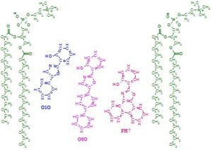
Localization and orientation of fluorescent probes O1O, O6O and PH7 in outer leaflet of phospholipid membranes. Two molecules of 1,2-dipalmitoyl-phosphatidylcholine are presented to show the location and orientation of the probes.